Non-Conjugated Double Bonds
The Author: Gerhard Knothe
The introduction of one double bond gives rise to several peaks in the NMR spectrum compared to the saturated chains in methyl stearate or stearic acid.
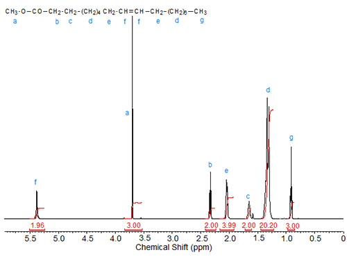
In methyl oleate (methyl 9(Z)-octadecenoate; Fig. 1), the following changes can be observed in comparison to methyl stearate and stearic acid: Two olefinic protons (integration value = 2) at about 5.3 ppm, four allylic protons (at C8 and C11) at about 2.05 ppm. The theoretical integration value of the strong CH2 peak decreases to 20.
Figure 1. 1H-NMR spectrum of methyl oleate.
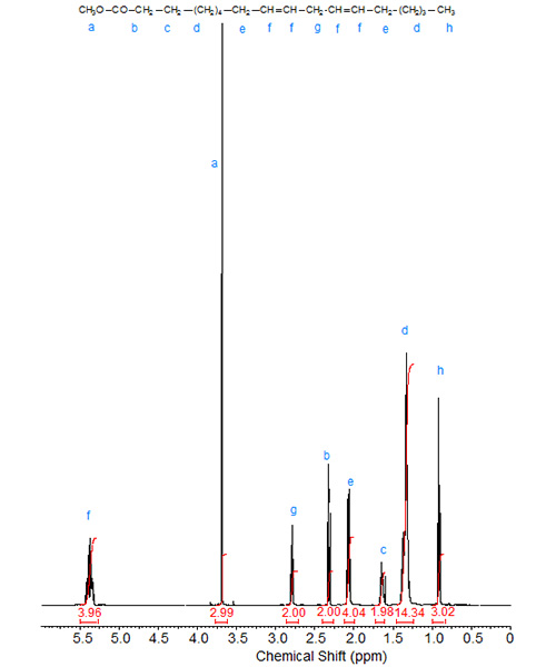
Introduction of a second double bond to give linoleic acid or methyl linoleate (methyl 9(Z),12(Z)-octadecadienoate; Fig. 2) gives rise to a peak at 2.8 ppm caused by the bis-allylic protons located at C11. The theoretical integration value of the olefinic protons increases to four while that of the large CH2 peak decreases further to 14.
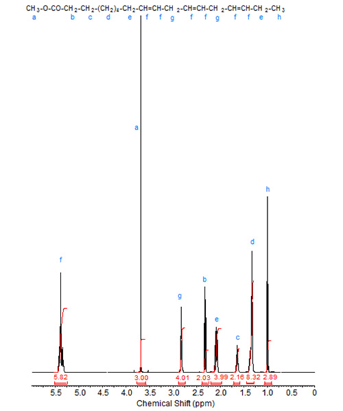
A third double bond in linolenic acid or methyl linoleate (methyl 9(Z),12(Z)-octadecatrienoate; Fig. 3) does not cause any new peaks compared to linoleic acid or methyl linoleate, only changes in the integration values. However, the triplet of the terminal methyl group (ω1) shifts downfield slightly, to about 0.95 ppm. This shift can be used for quantification purposes in mixtures as the terminal methyl triplet can be integrated separately from the corresponding signals of the other fatty acids.
Figure 3. 1H-NMR spectrum methyl α-linolenate.
"Migration" of the double bond leads to shift of signals, especially when the double bond approaches one of end of the chain. This effect was discovered in early NMR studies of a full series of cis-octadecenoic and some acetylenic fatty acids (Gunstone and Ismail, 1967). Prior work demonstrated this effect for double bonds near the terminal methyl group (Glass and Dutton, 1964; Storey 1960). The shifts of the olefinic protons do not differ significantly, if at all, for positions toward the middle of the chain (C8-C12 for a C18 chain). For determining such positions in a chain, a shift reagent [Eu(fod)3d30] was used (Frost and Sies, 1974; Bus and Frost, 1976). Similar work was carried out for unsaturated triacylglycerols using [Pr(fod)3] (Frost and Gunstone, 1975).
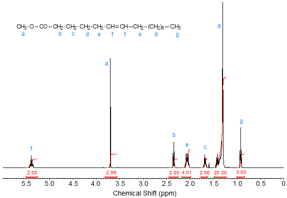
The 1H-NMR spectrum of the C18 fatty acid methyl ester methyl petroselinate (Fig. 4), in which the cis double bond is located at C6, however, shows minor changes in the chemical shifts compared to methyl oleate, such as the fine structure of the signals of the olefinic protons and a slight downfield shift of the triplet caused by the C2 protons.
Figure 4. 1H-NMR spectrum of methyl petroselinate.
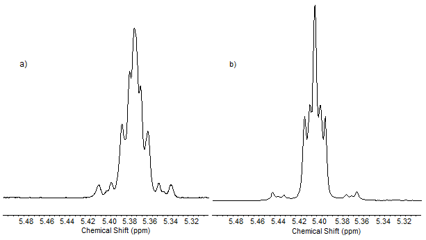
Double bond configuration affects the NMR signals of the unsaturated fatty compounds. The compounds whose spectra are depicted in Figures 1 to 4 all possess cis double bonds. Figure 5 shows the NMR spectrum of methyl elaidate, the Δ9 E-isomer of methyl oleate. Figure 6 is an enlargement of the signals of the olefinic protons of methyl oleate and methyl elaidate. The smaller coupling constants for cis (6-15 Hz) than for trans (11-18 Hz) are visible in the spacing of the peaks. Table 1 gives some characteristic shifts of unsaturated fatty compounds.
Figure 5. 1H-NMR spectrum of methyl elaidate.
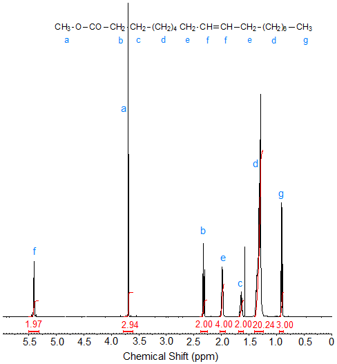
Figure 6. Expansion of the signals of the olefinic protons in the 1H-NMR spectra of (a) methyl oleate and (b) methyl elaidate.
| Table 1. Chemical shifts of cis-octadecenoic acids (Gunstone and Ismail, 1967; recalculated from τ values; 60 MHz NMR); solvent CCl4 | ||||
| Double bond position | —CH2—COOH | —CH2—CH=CH— | —CH=CH— | —CH3 |
|---|---|---|---|---|
| 2 | 2.65 (C2, C4) | 6.28, 6.10 (C3), 5.70 (C2) | 0.90 | |
| 3 | 3.01 | 2.08 | 5.52 | 0.91 |
| 4 | 2.30 | 2.10 | 5.37 | 0.91 |
| 5 | 2.10 | 5.36 | 0.90 | |
| 6 | 2.10 | 5.35 | 0.90 | |
| 7 | 2.10 | 5.34 | 0.90 | |
| 8 | 2.10 | 5.32 | 0.91 | |
| 9 | 2.10 | 5.32 | 0.91 | |
| 10 | 2.10 | 5.32 | 0.90 | |
| 11 | 2.10 | 5.32 | 0.91 | |
| 12 | 2.10 | 5.32 | 0.91 | |
| 13 | 2.10 | 5.31 | 0.91 | |
| 14 | 2.10 | 5.32 | 0.92 | |
| 15 | 2.10 | 5.31 | 0.96 | |
| 16 | 2.10 | 5.36 | 1.62 | |
| 17 | 2.15 | 4.95 (C18), 5.60 (C17) | ||
| Methyl oleate |
2.3 | 2.0 | 5.27 | 0.9 |
| Methyl elaidate |
2.3-2.4 | 2.15 | 5.40 | 0.95 |
| Table 2. Chemical shifts of cis- and trans-octadecenoic acids and esters (Frost and Gunstone, 1975; solvent, carbon tetrachloride (CCl4); 220 MHz). This paper also features a discussion of shielding and deshielding effects and depicts various peak patterns. | ||||||
| Double bond | —CH2—COOR | —CH2—CH=CH— | —CH=CH— | |||
|---|---|---|---|---|---|---|
| Acid | Ester | Acid | Ester | Acid | Ester | |
| 2 cis | - | - | 2.645 | 2.625 | 6.285 (C3), 5.735 (C2) | 6.145 (C3), 5.680 (C2) |
| 2 trans | - | - | 2.21 | 2.18 | 7.01 (C3), 5.75 (C2) | 6.85 (C3), 5.72 (C2) |
| 3 cis | - | 3.06 (C2) | - | 3.06 (C2), 2.03 (C5) | - | 5.51 |
| 3 trans | 2.995 (C2) | 2.930 (C2) | 2.015 (C5) | 2.015 (C5) | 5.50 | 5.47 |
| 4 cis | - | 2.27 (C2, C3) | - | 2.270 (C2, C3), 2.025 (C6) | - | 5.31 |
| 4 trans | 2.33 (C2, 3) | - | 2.33 (C2, 3), 1.95 (6) | - | 5.40 | - |
| 5 cis | 5.32 | |||||
| 5 trans | 2.28 | - | 1.945 (C7), 2.025 (C4) | - | 5.34 | - |
| 6 cis | 1.995 (C8), 2.04 (C5) | 1.990 (C8), 2.02 (C5) | 5.29 | |||
| 6 trans | 2.295 | 2.215 | 1.950 (C9), 1.99 (C5) | 1.950 (C9), 1.975 (C5) | ||
| 7 cis | 1.985 (C9), 2.01 (C6) | 1.980 (C9), 2.0005 (C6) | 5.28 | |||
| 7 trans | 2.290 | - | 1.935 (C9), 1.960 (C6) | - | ||
| 8 cis | 5.28 | |||||
| 8 trans | ||||||
Frost and Gunstone, 1975; Δ12-Δ15 determinable by effect on terminal methyl:
|
||||||
| Table 3. Chemical shifts in methyl cis,cis-octadecadienoates (Gunstone et al., 1969). Solvent CCl4. | |||
| Double bond position | —CH2—COOMe | —CH2—CH=CH— | —CH=CH— |
|---|---|---|---|
| 7, 15 | 1.96, 2.01 | 5.26 | |
| 8, 15 | 1.96, 2.01 | 5.26 | |
| 5, 12 | 1.96, 2.01 | 5.27 | |
| 9, 15 | 1.96, 2.01 | 5.26 | |
| 6, 12 | 1.99 | 5.28 | |
| 7, 12 | 1.97, 2.02 | 5.28 | |
| 6, 11 | 1.99, 2.04 | 5.30 | |
| 8, 12 | 2.03 | 5.29 | |
| 6, 10 | 2.02 | 5.29 | |
| 9, 12 | 1.99, 2.03 | 5.26 | |
| 6, 9 | 2.04 | 5.28 | |
| 10, 12 | 2.09, 2.16, 2.20 | 5.32 (C10, C13), 6.12 (C11, C12) | |
| 6, 8 | 2.1-2.5 | 5.1-5.9 | |
The differences in the coupling constants between cis and trans can be visualized by expanding the peaks of the olefinic protons:
The cis and trans isomers can be distinguished by the coupling constants as well as by some chemical shifts. For example, the signals of the allylic protons in elaidic acid are shifted slightly upfield (by about 0.05 ppm) compared to oleic acid. On the other hand, the signals of the olefinic protons of elaidic acid are slightly downfield (by about 0.03 ppm) compared to oleic acid. Thus, the shift difference between the peaks of the allylic and the olefinic protons is suitable for distinguishing cis/trans isomers, with the difference being greater for trans. This effect has been discussed in the literature (Frost and Gunstone, 1975), where also the δ-values for each methylene group were calculated.
Gunstone and Jacobsberg (1972) gave the chemical shifts of all isomers of 9,12-diunsaturated C18 acids. See Table 4 for the values of those containing only double bonds.
| Table 4. Chemical shifts in 9,12-dienoic C18 fatty acids (Gunstone and Jacobsberg, 1972; solvent probably CCl4)a | |||
| Isomer | —CH=CH— | =HC—CH2—CH= | —CH=CH—CH2— |
|---|---|---|---|
| 9c,12c | 5.27 | 2.71 | 1.99 |
| 5.25b | 2.68 b | 2.10-2.20b | |
| 9c,12t | 5.31 | 2.67 | 1.97 |
| 9t,12c | 5.31 | 2.68 | 1.97 |
| 9t,12t | 5.32 | 2.62 | 1.96 |
|
aValues originally reported on the τ scale, converted to δ scale here. |
|||
Literature:
- Bus, J. and Frost, D.J. Determination of the positions of double bonds in unsaturated fatty acids by 13C and proton NMR spectrometry. Lipids, [Invited Lect. Symp. Int. Congr. Fat Res. (1974)] Vol. 2, 343-350 (pub. 1976).
- Frost, D.J. and Gunstone, F.D. The PMR analysis of non-conjugated alkenoic and alkynoic acids and esters. Chem. Phys. Lipids, 15, 53-85 (1975).
- Frost, D.J. and Sies, I. PMR Analysis of alkenoic esters using shift reagents. Chem. Phys. Lipids, 13, 173-177 (1974).
- Glass, C.A. and Dutton, H.J. Determination of beta-olefinic methyl groups in esters of fatty acids by nuclear magnetic resonance. Anal. Chem., 36, 2401-2404 (1964).
- Gunstone, F.D. 1H- and 13C-NMR spectra of six n-3 polyene esters. Chem. Phys. Lipids, 56, 227-229 (1990).
- Gunstone, F.D. and Ismail, I.A. Fatty acids, Part 15. Nuclear magnetic resonance spectra of the cis octadecenoic acids and of some acetylenic acids. Chem. Phys. Lipids, 1, 337-340 (1967).
- Gunstone, F.D., Lie Ken Jie, M. and Wall, R.T. Fatty acids. Part 23. Nuclear magnetic resonance spectra of some octadecadiynoic acids and of some methyl cis,cis- and trans,trans-octadecadienoates. Chem. Phys. Lipids, 3, 297-303 (1969).
- Gunstone, F.D. and Jacobsberg, F.R. Fatty acids, Part 36. The synthesis, silver ion chromatographic, and NMR spectroscopic properties of the nine 9,12-diunsaturated n-C18 acids. Chem. Phys. Lipids, 9, 112-122 (1972).
- Hashimoto, T., Nukada, K., Shiina, H. and Tsuchiya, T. On the structure of highly unsaturated fatty acids of fish oils by high resolution nuclear magnetic resonance spectral analysis. J. Am. Oil Chem. Soc., 40, 124-128 (1963).
- Kannan, R., Subbaram, M.R. and Achaya, K.T. NMR studies of some oxygenated, halogenated, and sulphur-containing fatty acids and their derivatives. Fette Seifen Anstrichm., 76, 344-350 (1974).
- Lie Ken Jie, M.S.F. and Lam, C.C. 1H-Nuclear magnetic resonance spectroscopic studies of saturated, acetylenic and ethylene triacylglycerols. Chem. Phys. Lipids, 77, 155-171 (1995).
- Sacchi, R., Medina, I., Aubourg, S.P., Addeo, F. and Paolillo, L. Proton nuclear magnetic resonance rapid and structure-specific determination of ω-3 polyunsaturated fatty acids in fish lipids. J. Am. Oil Chem. Soc., 70, 225-228 (1993).
- Storey, W.H. Fatty acids analysis by high resolution nuclear spin resonance. A preliminary evaluation. J. Am. Oil Chem. Soc., 37, 676-678 (1960).
In This Section
- Introduction of NMR
- Saturated Fatty Acids and Methyl Esters
- Alkyl Esters Other than Methyl
- Glycerol Esters
- Non-Conjugated Double Bonds
- Conjugated Linoleic Acid (CLA)
- Acetylenic Fatty Acids and Derivatives
- Branched-Chain and Cyclic Fatty Acids
- Epoxy Fatty Acids
- Hydroxy and Hydroperoxy Fatty Acids
- Oxo Fatty Acids
- Fatty Alcohols
- Some Miscellaneous Fatty Acids
- Quantification by 1H-NMR
- The NMR Spectrum
- Alkanoic Acids
- Monoenoic Acids
- Polyunsaturated Fatty Acids
- Non-Methylene-Interrupted Polyenoic Fatty Acids
- Acids with conjugated unsaturation
- Acetylenic and Allenic Acids and Esters
- Branched-Chain and Cyclic Fatty Acids
- Cyclic Fatty Acids
- Epoxides and Acyclic Ethers
- Hydroxy and Hydroperoxy Acids
- Oxo (Keto) Acids
- Acids, Esters (Alkyl, Glycerol, Waxes), Alcohols and Acetates, Amides, and Nitriles
- Esters of Glycerol and Other Polyhydric Alcohols
- Oils and Fats
- Regiospecific Analysis of Triacylglycerols